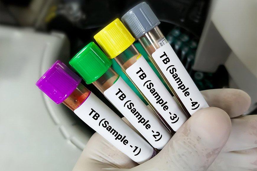
What are tumor markers and what types are there?
What are tumor markers and what types are there? Belgrade. TOP PRICE✓ Tumor diagnostics✓ Precise testing✓ Early detection of cancer✓ Laboratory diagnostics of tumors✓
What are tumor markers and what types are there?
Detailed laboratory analyses are not unfamiliar when we need insight into a wide range of conditions affecting our internal organs and the processes occurring within them.
Laboratory analyses, especially blood samples, are now routinely performed and take very little time, with discomfort minimized to a minimum.
Specific blood analyses are conducted by specialized laboratory staff, and the most advanced equipment processes the samples and prepares reliable results. Often, it is possible to send results to other countries for analysis and thus obtain a more comprehensive view of health status or get a second opinion.
One of these specific analyses that highlight the importance of quality technology and medical knowledge is blood tests searching for tumor markers.
What are tumor markers?
Tumor markers or tumor markers are substances produced by diseased or abnormal cells in an organ, as well as by healthy, normal cells—but in much smaller quantities, usually due to benign disorders in organ function.
These substances are produced either by the cancer itself or by the patient's body in response to the presence of cancer. Although tumor markers are found within cells, they also circulate in the blood.
Let’s better understand how tumors affect our organs.
A tumor can originate from any cell in the body that undergoes abnormal growth and proliferation. Any cell in the body can undergo such proliferation, but what is characteristic of tumor cells is:
- Autonomous growth
- Undifferentiated and primitive structure
- Infiltrative growth
- Altered metabolism
- Ability to metastasize
What conditions lead to the development of tumors?
Disrupted activities of proteins crucial in regulating all cellular processes, such as cell signaling and apoptosis control, lead to disease.
When the regulation of proliferation (normal differentiation and cell multiplication) in multicellular organisms (humans, animals, etc.) is disrupted, it results in uncontrolled division and spread of the “problematic” cell throughout an organ, which impairs the normal function of healthy cells.
What are tumor markers used for?
Tumor markers are generally not used for diagnosis and screening. The presence of cancer can only be confirmed by biopsy. However, tumor markers play a significant role in detecting the source of primary cancer in patients without a prior history, as well as in early detection of tumor recurrence in individuals who have already battled the condition.
Tumor markers are also used to assist in diagnosis, disease staging, predicting disease course, and planning and monitoring therapy, i.e., predicting the response of squamous cells to treatment.
What makes an ideal tumor marker? What is actually involved?
- A tumor marker that is specific enough to detect and indicate the exact type of cancer is considered an “ideal” situation.
- It must also have sufficient sensitivity—meaning that it should be detectable in early stages of the disease—to allow for timely detection (so that tumor markers can identify it).
- The amount of substance secreted should be proportional to the size of the tumor cell that secretes it, to provide insight into its development.
Unfortunately, no ideal tumor marker exists.
Although theoretically possible to develop such a marker, none known so far is 100% specific or 100% sensitive (as it cannot be detected in all disease stages, particularly in the early stages).
Despite these “shortcomings,” tumor markers are routinely determined in laboratory practice, and their usefulness in detecting and monitoring diseases cannot be denied.
What else should be known about determining tumor markers?
When discussing tumor markers, we are referring to substances that are most commonly produced by malignant cells, although they can also be produced by healthy cells as a response to tumor tissue.
Increased levels or elevated tumor markers must be persistent in repeated blood analyses—to establish a cancer diagnosis in general.
Serial determination of tumor markers ensures detection of false-positive values due to transient increases. Whenever possible, all repetitions should be performed by the same laboratory using the same test.
Individual values are not very significant as various non-malignant conditions can cause elevated levels of tumor markers, but these increases are usually transient.
Tumor markers used in clinical practice
Tumor markers can be sought in various tissues and samples; these can include enzymes, specific proteins, different antigens, hormones, and specific receptors, etc.
Elevated tumor markers can be sampled from blood, urine, stool, but also from tumor tissues themselves, as well as from various bodily fluids in patients with malignant diseases.
Over 100 different tumor markers have been discovered today, but only about twenty are currently used in laboratory practice. Some tumor markers are associated with a single type of malignancy, while others can be associated with multiple types of tumor tissue.
-
Afp tumor markers – liver and (rarely) reproductive organs
In healthy adults, AFP tumor markers are present in minimal amounts or not at all. However, malignant liver cancer cells, embryonal tumors of the testes, ovaries, and uterus produce AFP (alpha-fetoprotein) intensely.
The greatest diagnostic value of AFP is as a tumor marker for the liver, confirming primary liver cancer, and its indicators are also significant in therapy and post-intervention for this organ.
-
β-2-microglobulin (β-2MG) – tumor markers for kidneys and kidney function
The level of β-2MG in the kidneys of healthy individuals is usually constant as it is normally broken down, filtered, and eventually excreted—while in impaired kidney function, the level of β-2MG increases.
-
β-human chorionic gonadotropin (β-HCG) – tumor markers for ovaries and uterus
These tumor markers are particularly significant in diagnosing choriocarcinoma—the most aggressive cancer in the human body. It is intensely produced in malignant teratomas of the uterus, ovaries, and sometimes the testes.
β-HCG is normally produced by the placenta from the 9th day of pregnancy onward.
A tumor marker known as CA-125 indicates the production of large amounts of cancerous fluid in the ovaries.
-
CEA tumor marker values for pancreas and colon – carcinoembryonic antigen
Carcinoembryonic antigens are physiologically produced in fetal tissue and are present in high quantities in fetal blood. After birth, the level of CEA tumor markers decreases.
In healthy adults, it is absent or present in very low quantities. If the CEA marker is elevated in the blood, it is significant for confirming malignancy of the pancreas, biliary tract, and colon.
Elevated CEA markers may also appear in the type CA50 in patients with benign pancreatic diseases and liver cirrhosis. It is used for monitoring patients with pancreatic cancer and in analyses required for organ transplantation preparations.
-
CA 15-3 – tumor markers for breast cancer
Elevated levels of CA 15-3 are an indicator of breast cancer. It is particularly useful in monitoring the course of the disease and therapy effectiveness in patients with diagnosed malignancies.
While the CA 15-3 marker is often elevated in advanced stages of breast cancer, it is generally less effective for early detection and diagnosis.
-
CA 19-9 – tumor markers for pancreas
CA 19-9 tumor marker is typically used in assessing the presence of malignancy in the pancreas, although it can also be associated with cancers of the liver and bile ducts. It helps in detecting tumor recurrence and in monitoring therapy progress.
CA 19-9 marker is valuable in distinguishing pancreatic cancer from other digestive system malignancies.
In summary, tumor markers represent valuable tools in detecting the presence of cancer, monitoring its course, and evaluating therapeutic responses.
Despite their limitations, they are indispensable in modern diagnostics and patient management, providing critical insights into disease status and treatment effectiveness.
How to Contact Us?
For more information about our services or if you have any questions, feel free to visit our CONTACT page and get in touch with us. We’re here to help!